

Gallery
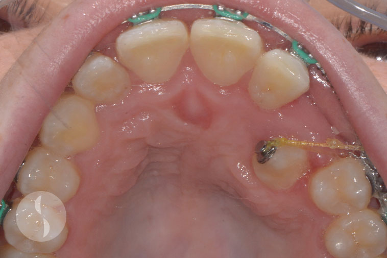
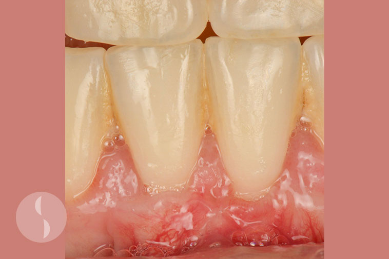
Periodontitis (Non Surgical)
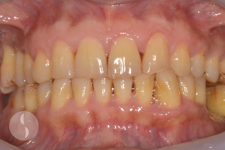
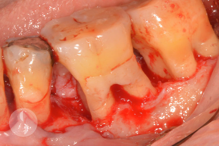
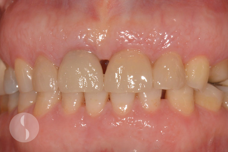
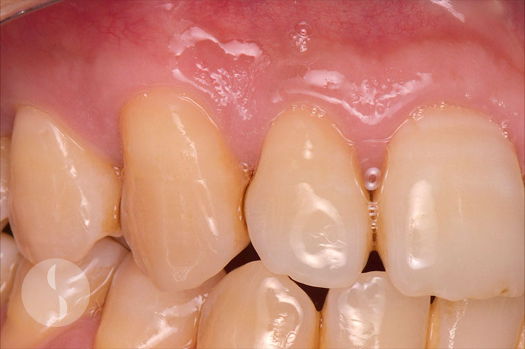
Case 1
Periodontitis
Treated non-surgically
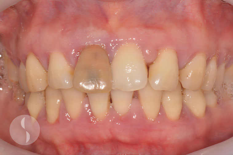
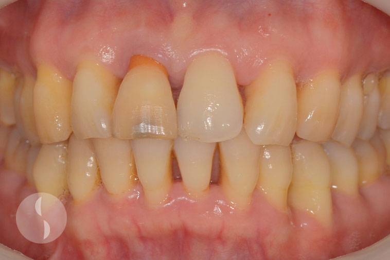
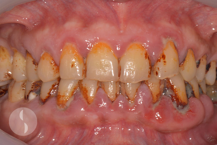
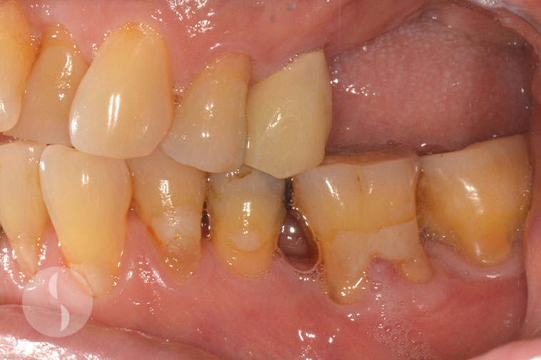
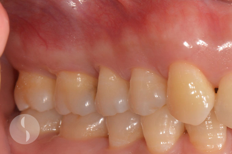
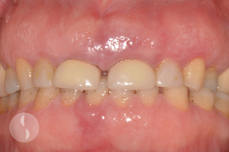
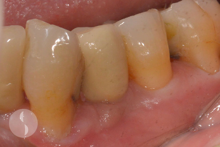
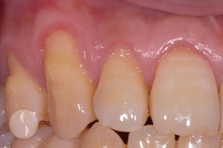
Case 2
Periodontitis
Treated non-surgically
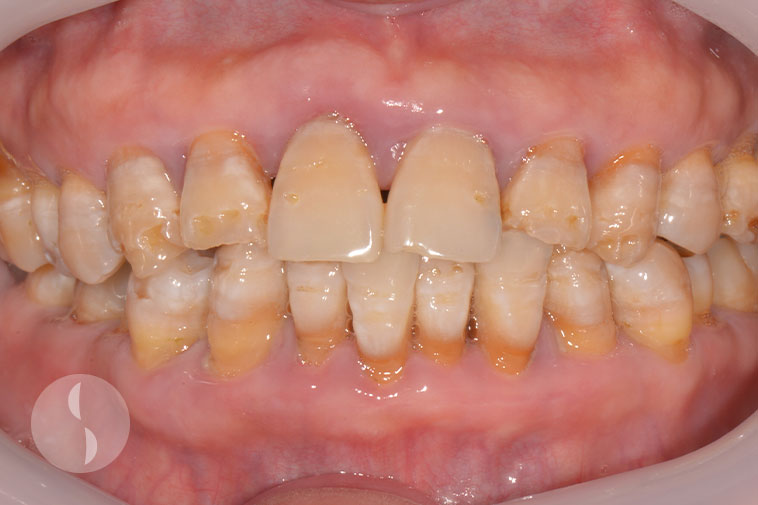
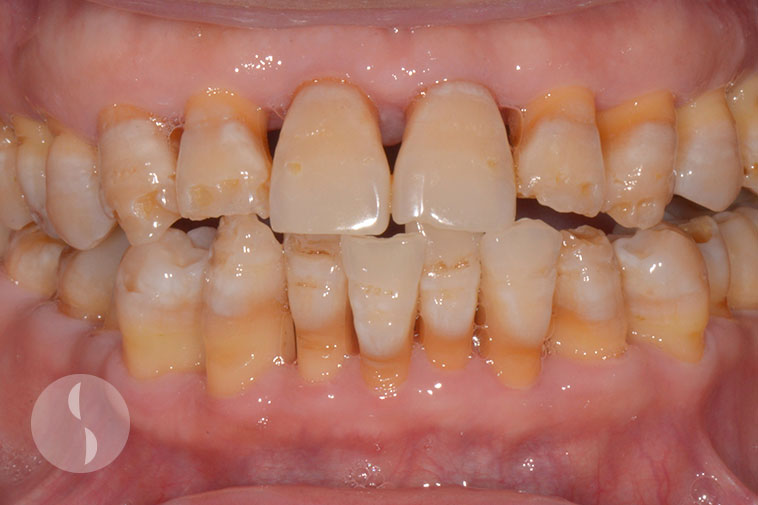
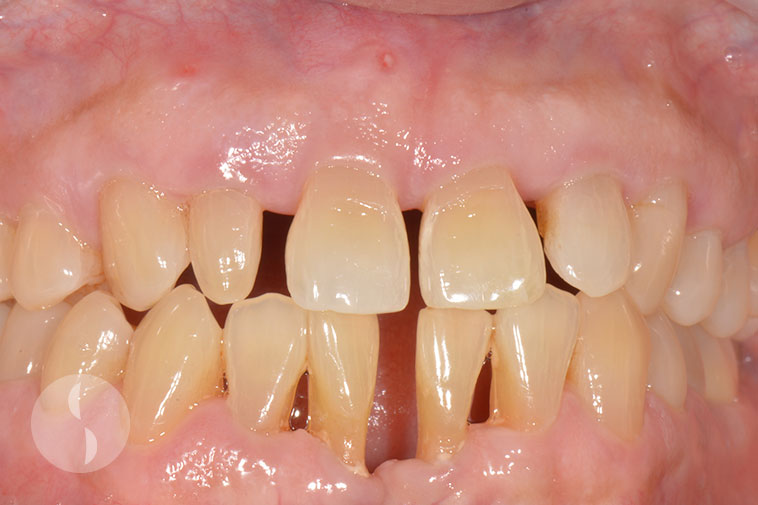
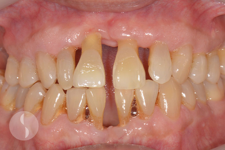
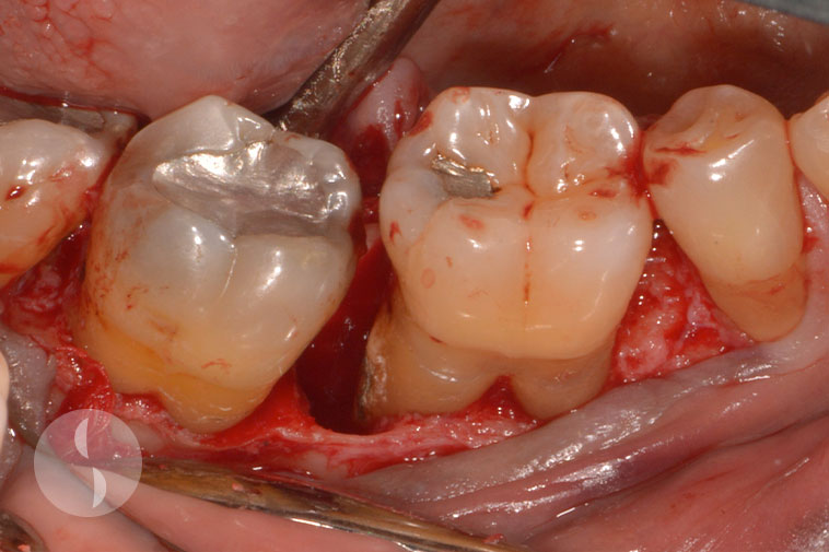
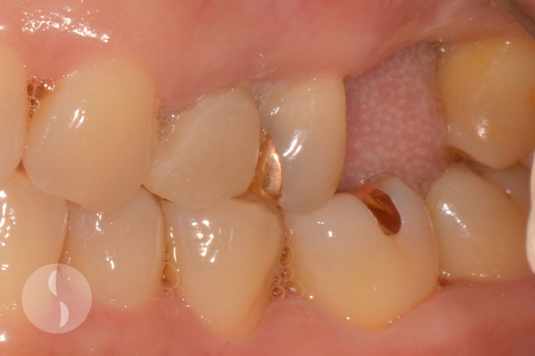
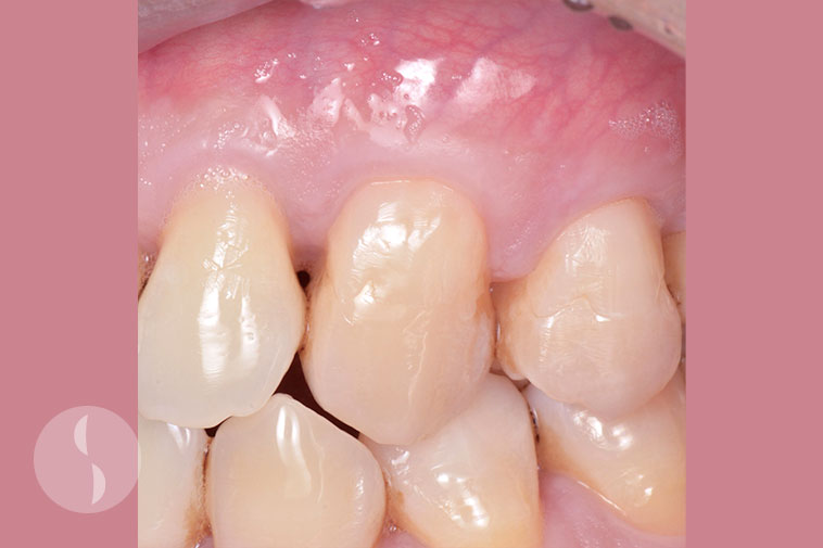
Case 3
Treated non-surgically
Periodontitis
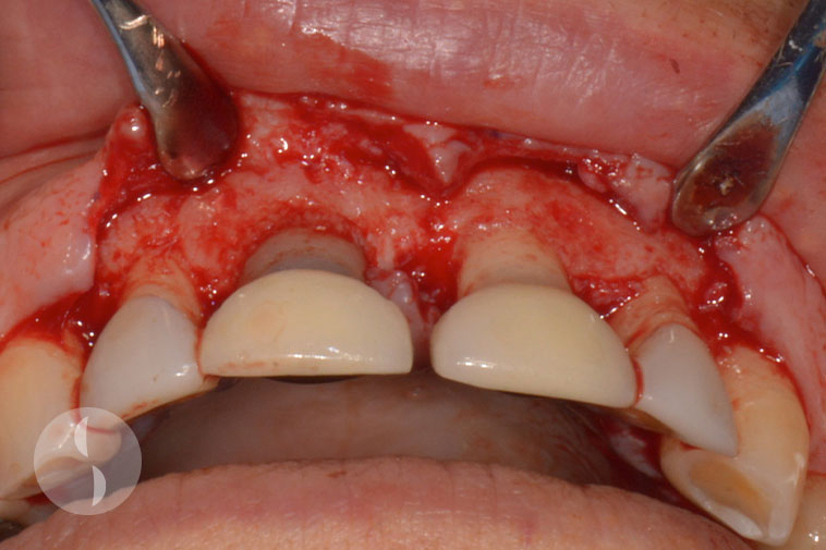
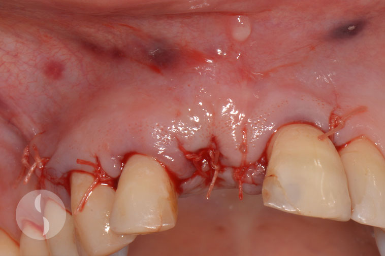
Periodontal Surgery
Case 1
Pre-operative
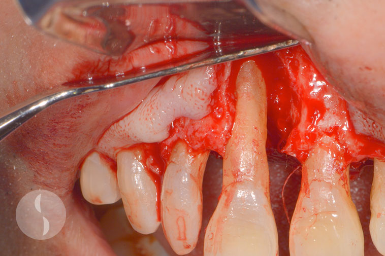
Large intrabony defect
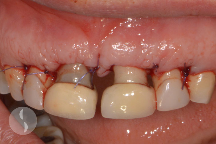
Tooth still maintained and functioning 1 year following surgery
Case 2
Surgical periodontal access surgery
Immediately following surgery
Periodontal health maintained at 6 months following surgery
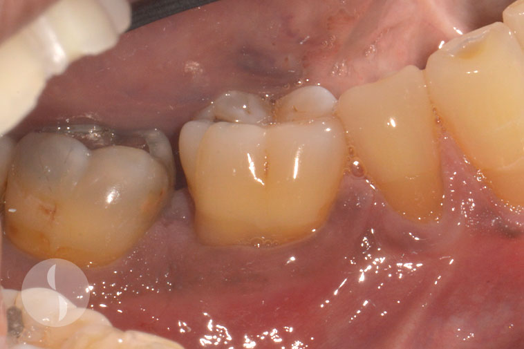
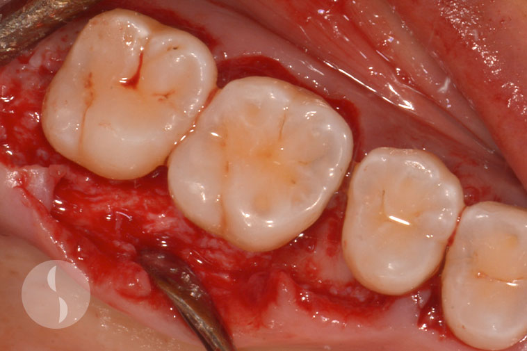
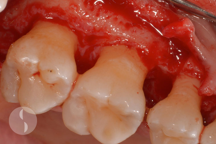
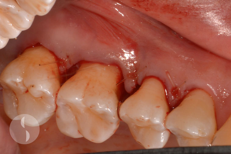
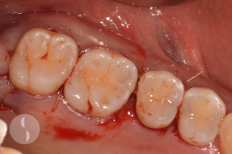
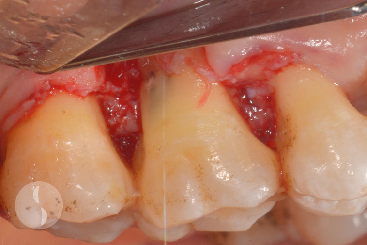
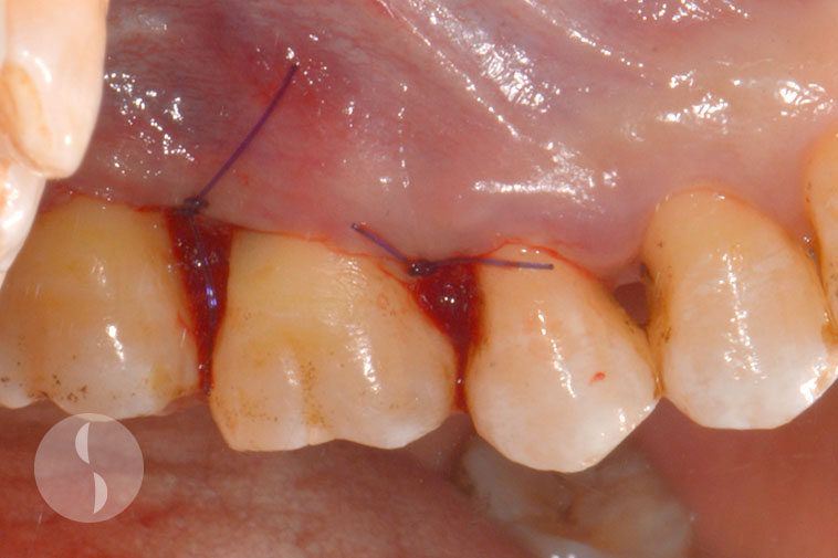
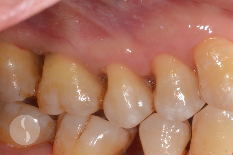
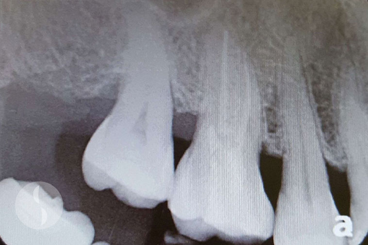
Case 3
Periodontal surgery for furcation and intrabony defects
Periodontal health maintained at 3 months following surgery
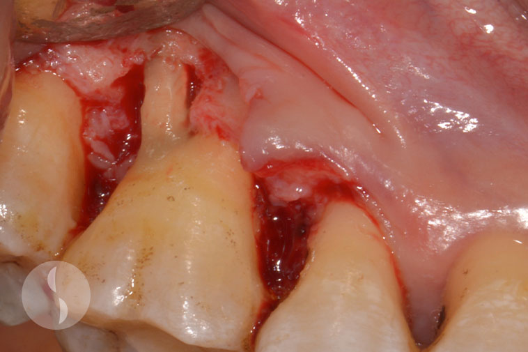
Case 4
Intrabony defect – 3 walled
Periodontal surgery providing access to debridement
Periodontal health achieved at 3 months following surgery
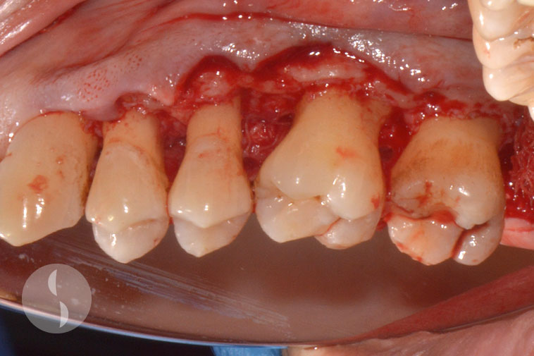
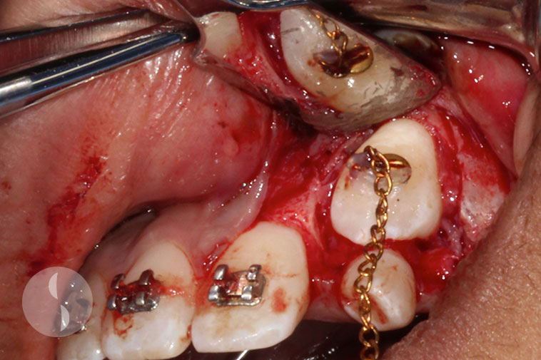
Case 5
Periodontal access surgery
Periodontal access for root decontamination
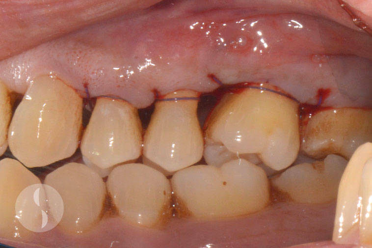
Immediate post operative
Periodontal health maintained 1 year following surgery
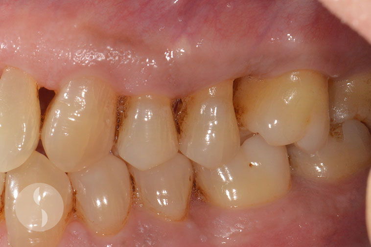
Immediate post operative
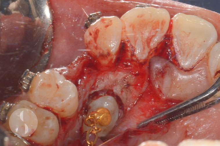
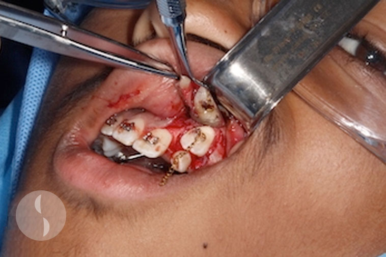
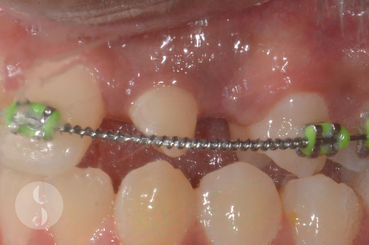
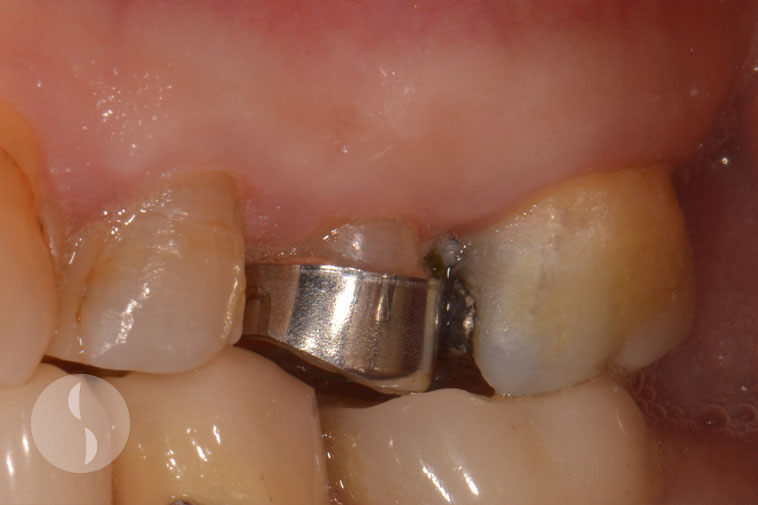
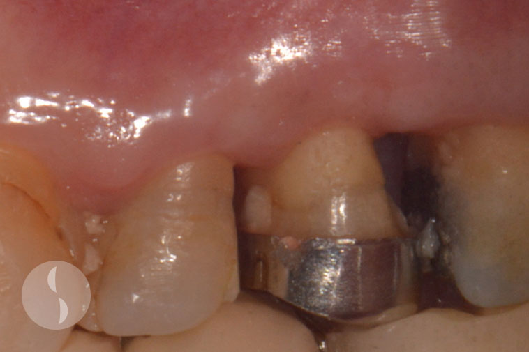
Root Resection
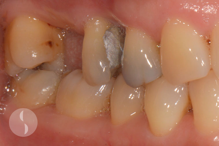
Case 1
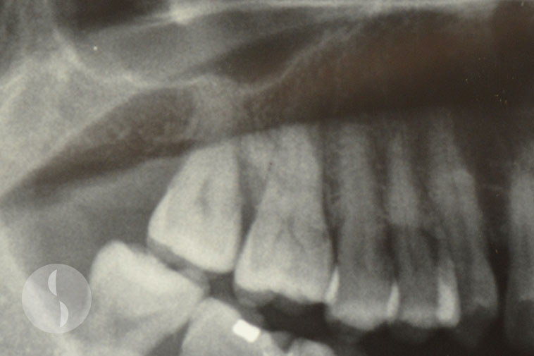
Pre-operative radiograph showing 16 distal furcation
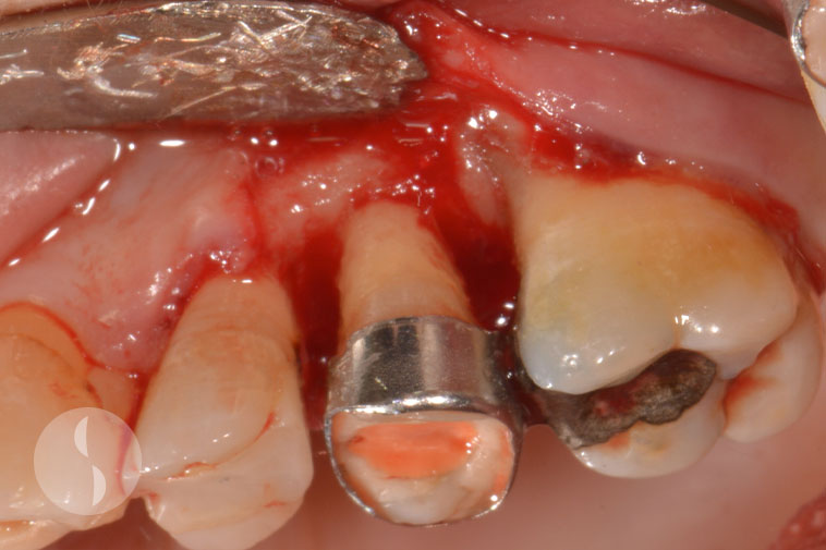
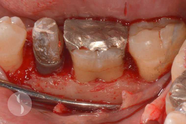
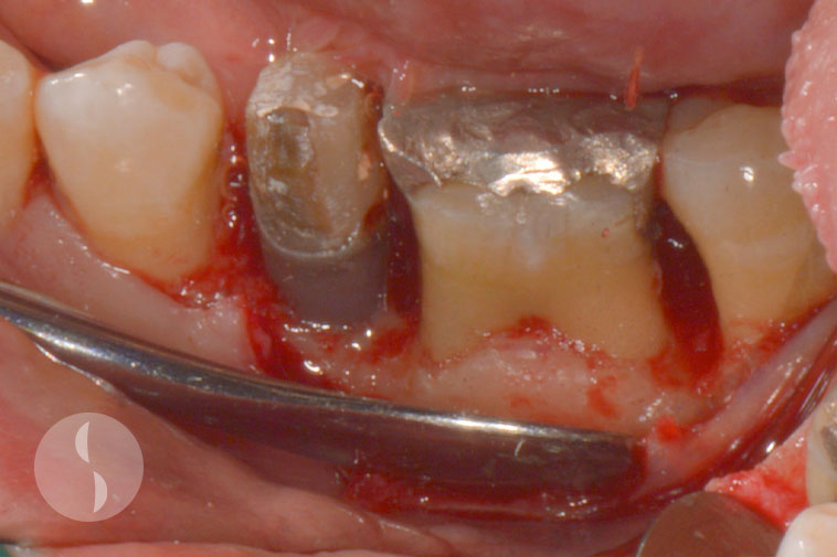
16 buccal bone removed around disto-buccal root
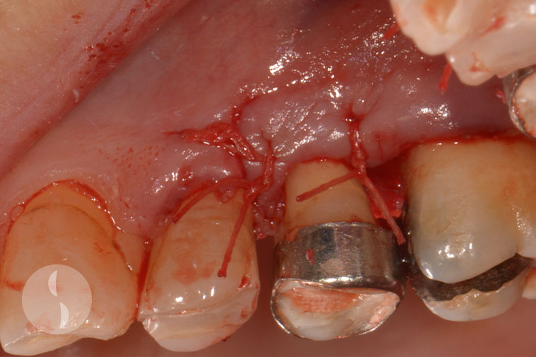
16 disto-buccal root resected
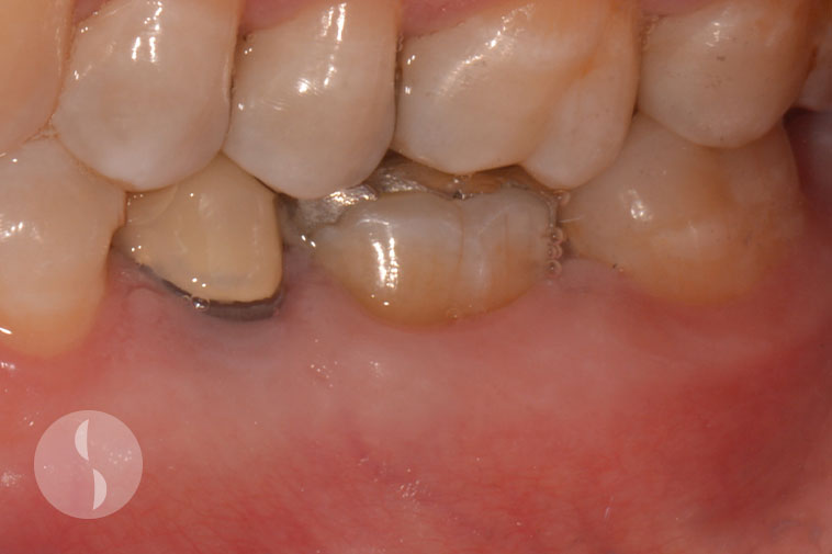
16 immediately post operative
Periodontal health maintained at 1 year following surgery
Post operative radiography 1 year following surgery
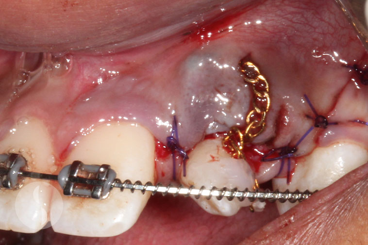
Canine Exposures
Case 1
13 canine palatal impaction
13 ‘closed’ exposure
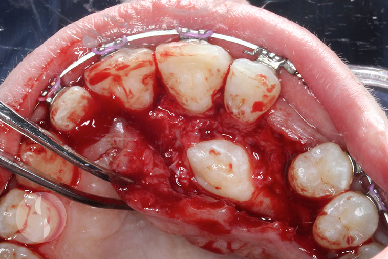
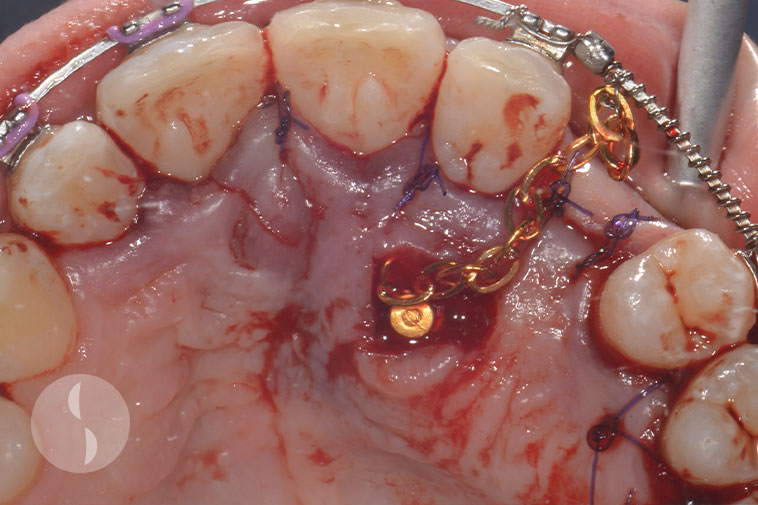
Case 2
Impacted canine 23
Exposure surgery
‘Closed’ canine exposure immediately post surgery
Case 3
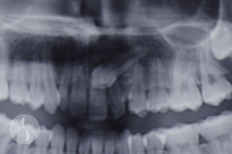
Pre-operative radiograph showing impacted canine 23
Palatal exposure of canine 23
‘Open’ canine exposure immediately post surgery
6 months post surgery
Crown Lengthening
Case 1
Pre-operative situation showing shortened clinical crowns of central incisors
Crown lengthening surgery
Immediately post surgery
Final result with incisors restored with veneers
Case 2
Pre-operative – tooth 25 with inadequate ferrule
Crown lengthening surgery on 25
Immediately following surgery
Increased ferrule at 3 month post surgery
Case 3
Tooth 36 inadequate ferrule for full coverage crown
Crown lengthening surgery 36
Crown lengthening surgery involving ostectomy
Increased clinical crown height at 8 weeks following surgery
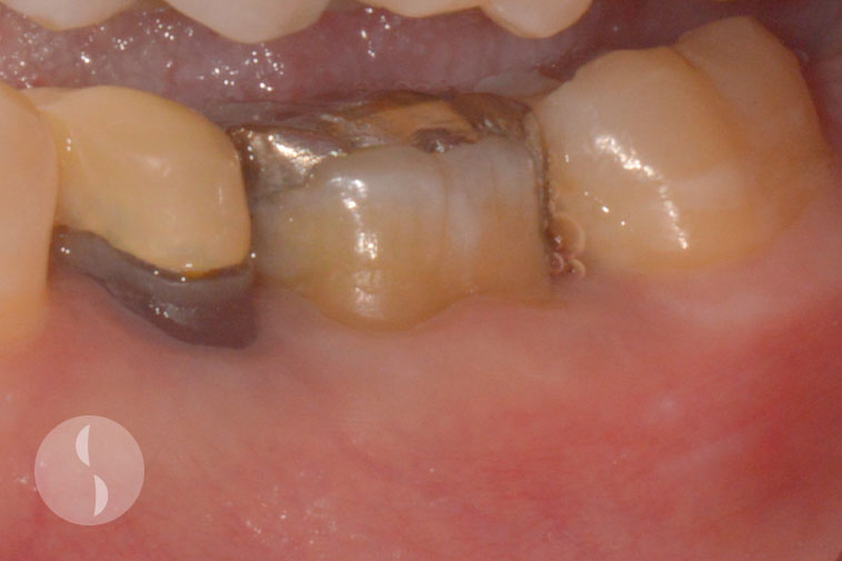
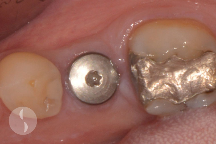
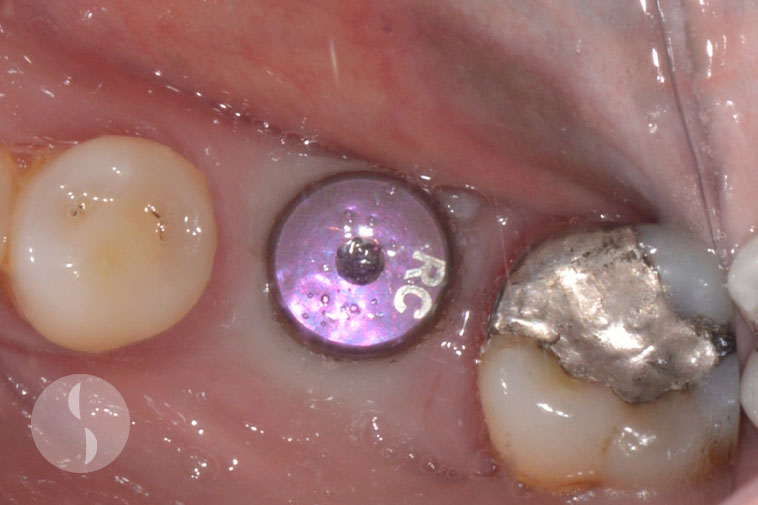
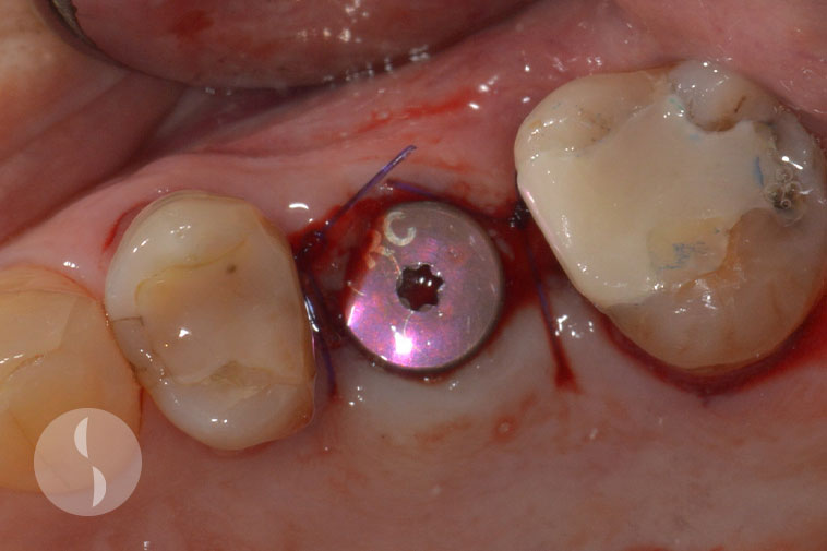
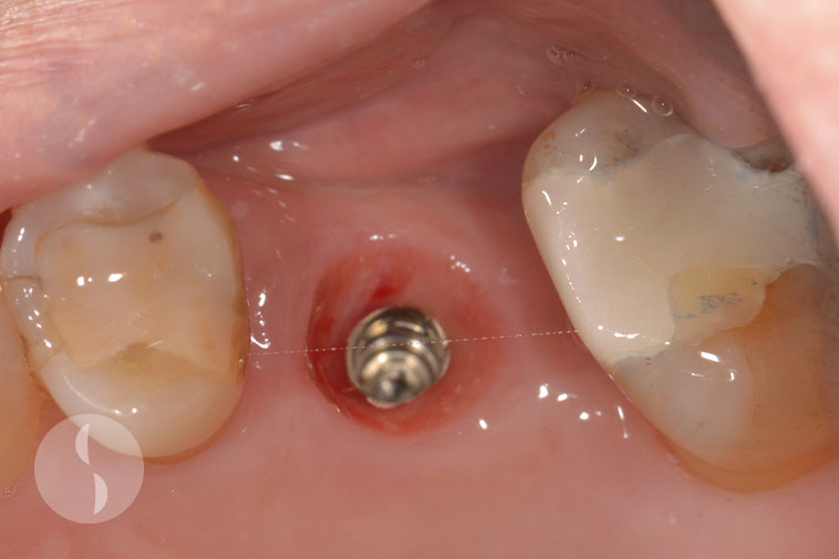
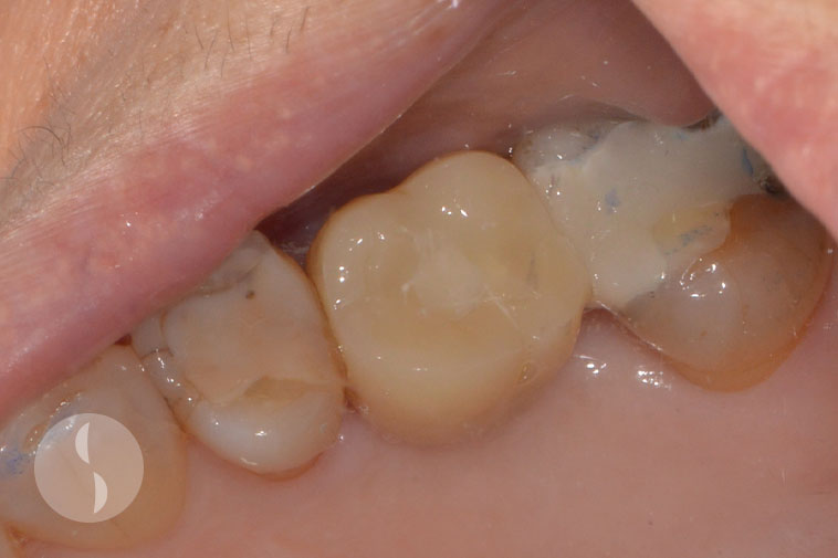
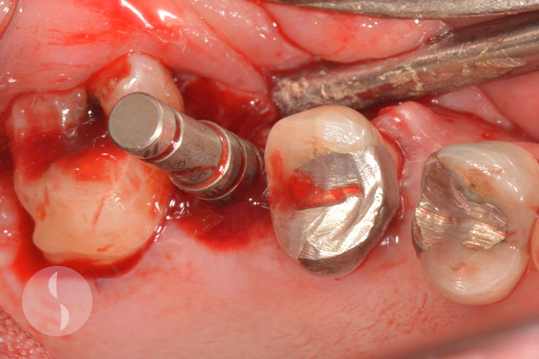
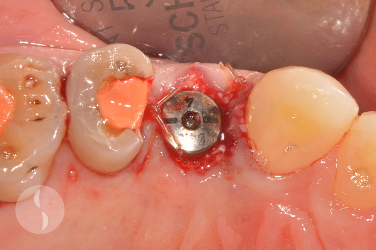
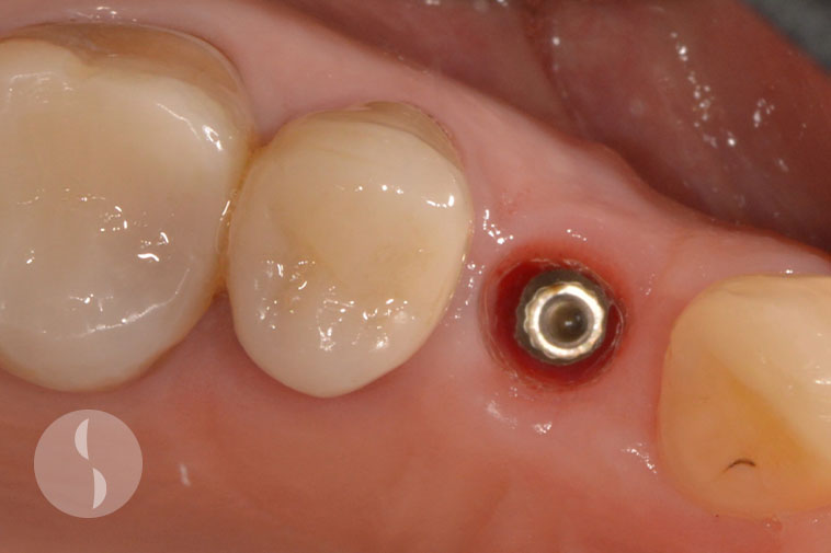
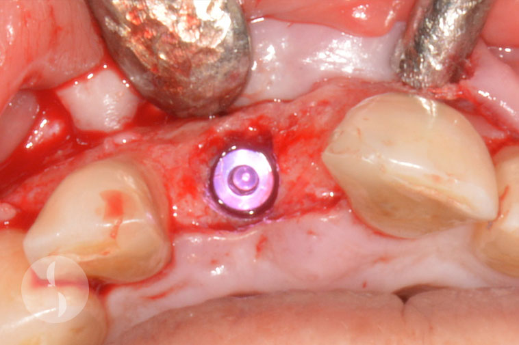
Standard Implant
Case 1
Excellent soft tissue healing at 8 weeks following surgery

Implant placed in prosthetically determined position
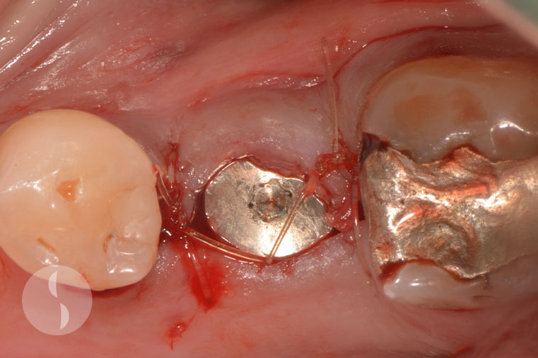
Semi-submerged technique for single stage implant surgery
Excellent soft tissue healing at 8 weeks following surgery
Case 2
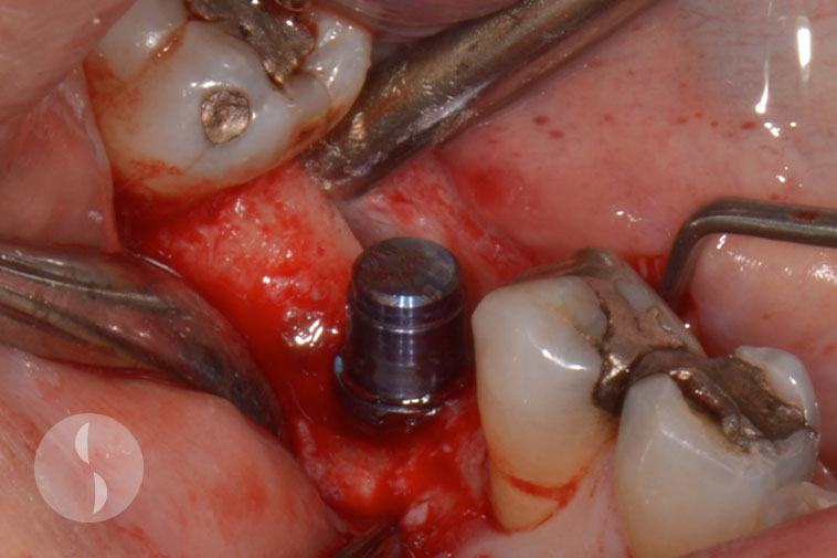
Implant surgery to replace 36
Implant placed with single stage technique
Excellent soft tissue healing at 8 weeks following surgery
Case 3
Direction indicator during implant surgery at 36
Immediately post operative showing implant placed as single stage
Excellent soft tissue healing 3 months following surgery
Case 4
Direction indicator during implant surgery
Single stage surgery for 26 implant
Healthy peri-implant mucosa
Provisional crown on 26 implant
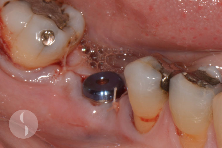
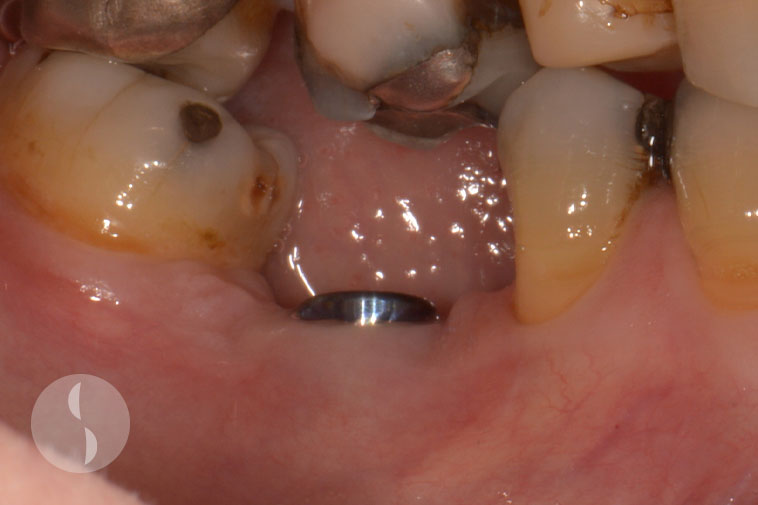
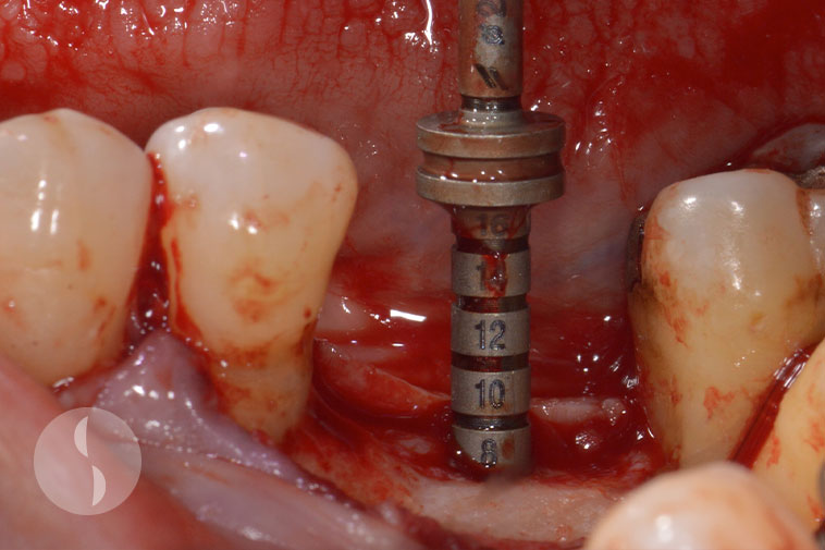
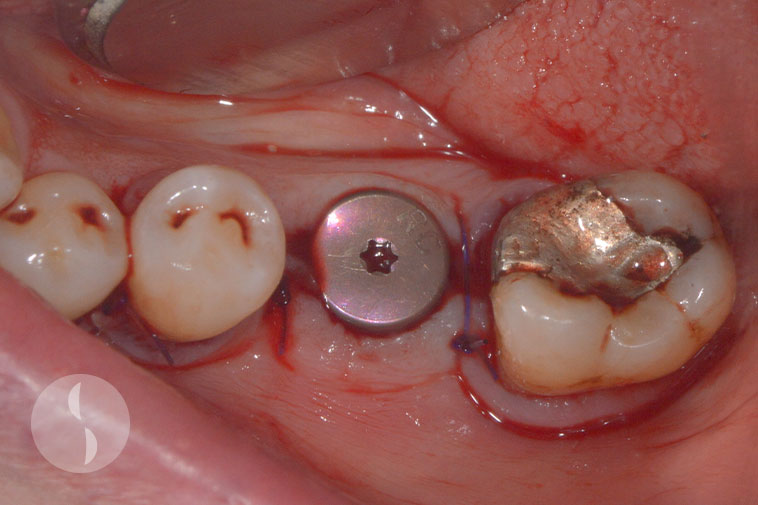
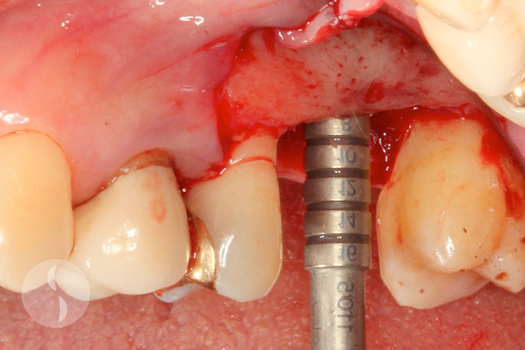
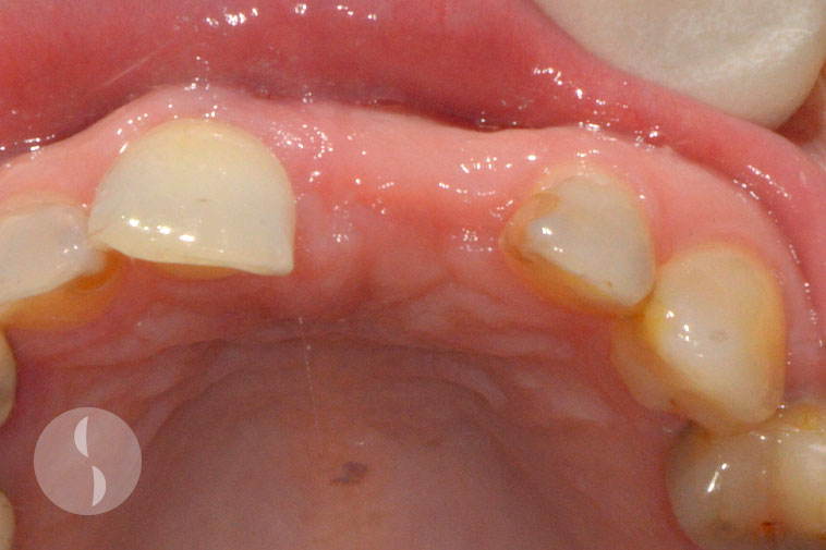
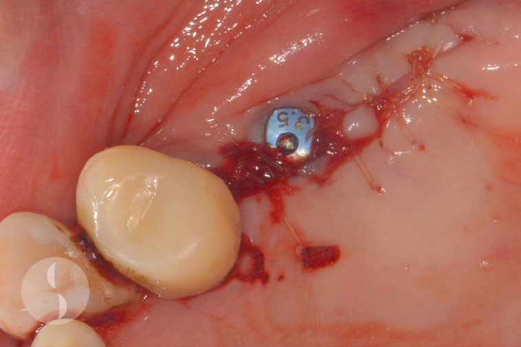
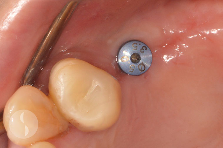
Case 5
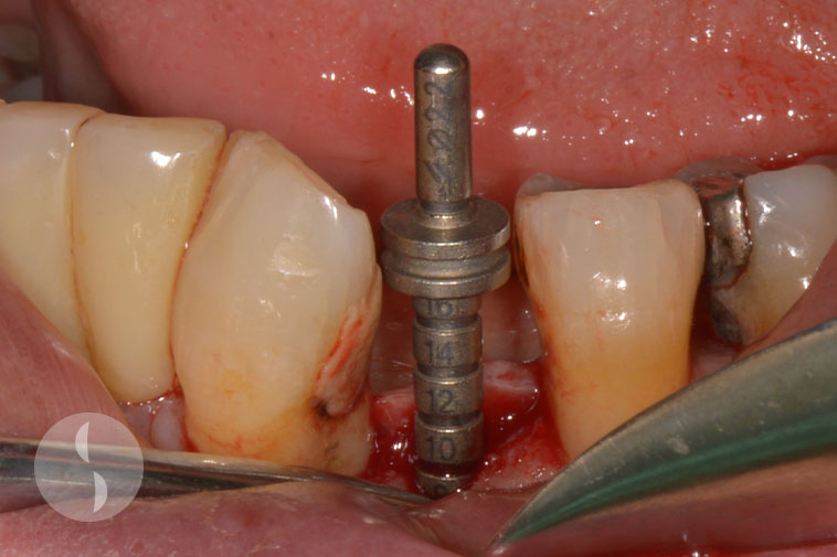
Narrow space for 34 implant
Implant placed in prosthetically determined position
Excellent soft tissue healing at 8 weeks following surgery
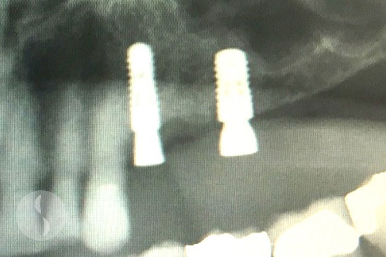
Follow up at 1 year following surgery
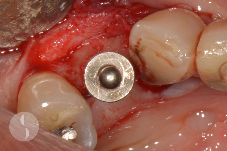
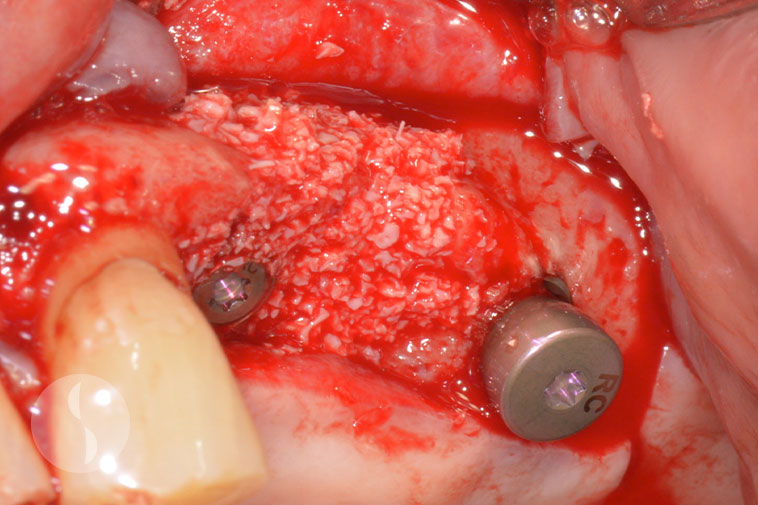
Case 6
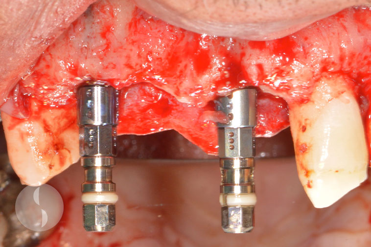
Pre-operative situation showing missing 26
Pre-operative situation showing missing 16
Direction indicator showing ideal positioning during implant surgery for 26
Direction indicator showing ideal positioning during implant surgery for 16
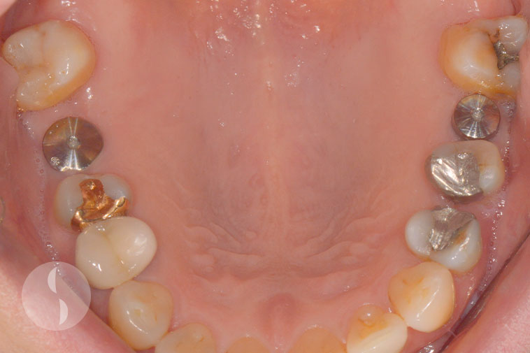
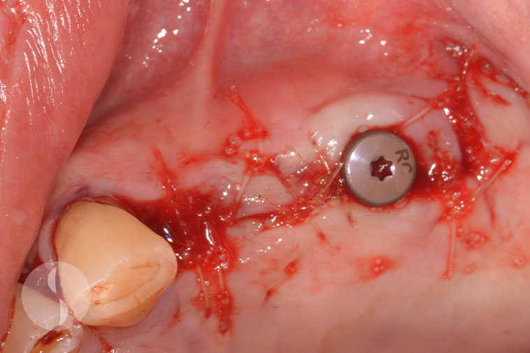
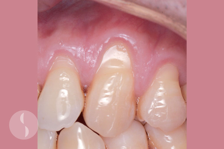
Excellent soft tissue healing at 8 weeks following surgery
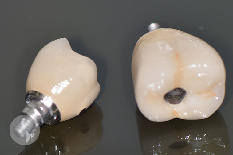
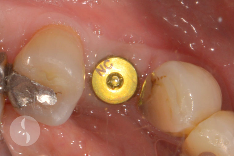
Screw retained crowns with ideal emergence profile

Issue of screw retained crowns for implants at 16 and 26
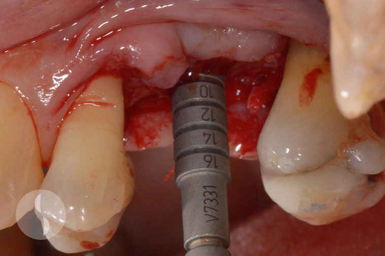
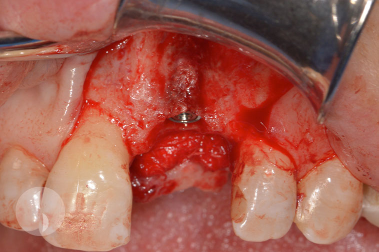
Complex Implant Surgery
Case 1
Narrow ridge width at implant site for 11
Ridge split in preparation for implant placement
Implant placed with crestal bone preserved on buccal aspect
Excellent integration at 3 months following surgery
Provisional crown at 11 Implant
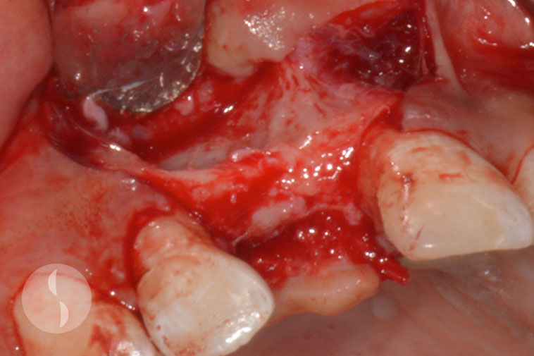
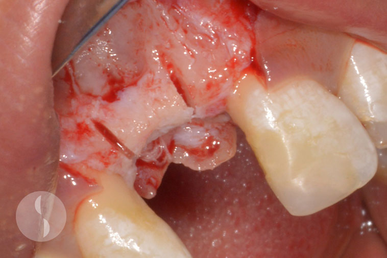
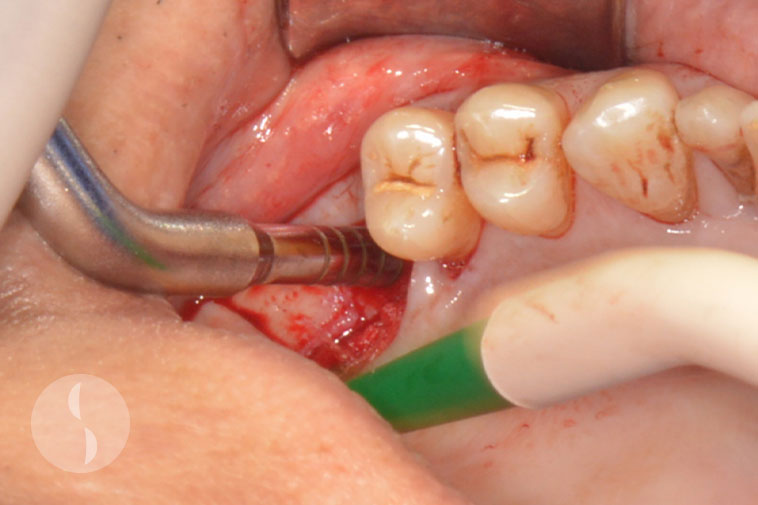
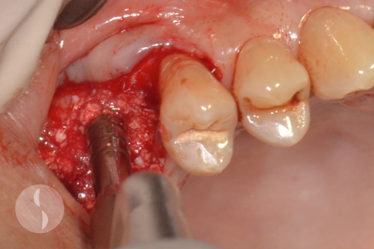
Case 2
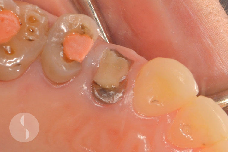
Unrestoratble 14 requiring extraction
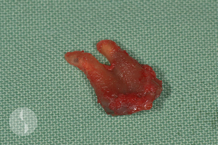
Extraction of 14
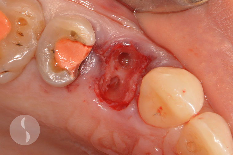
Socket 14 debrided
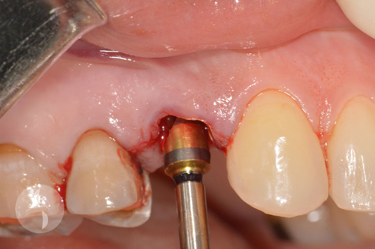
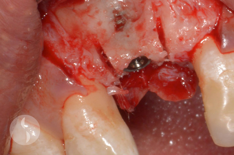
Immediate implant placement (flapless)
Immediate implant with grafting
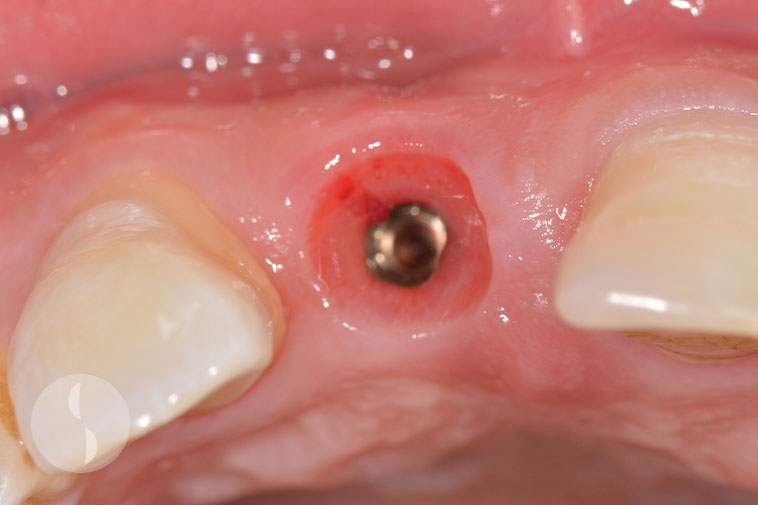
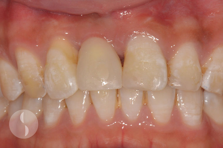
Excellent soft tissue integration at 8 weeks
Case 3
Preoperative situation showing missing 21 and flattening of buccal ridge
Implant placed with bone condensing technique to preserve buccal plate
Additional grafting (GBR) during implant placement
Placement of collagenous membrane for GBR technique
Case 4
Surgical stent showing ideal prosthetic planning
Implants placed according to prosthetically determined positions
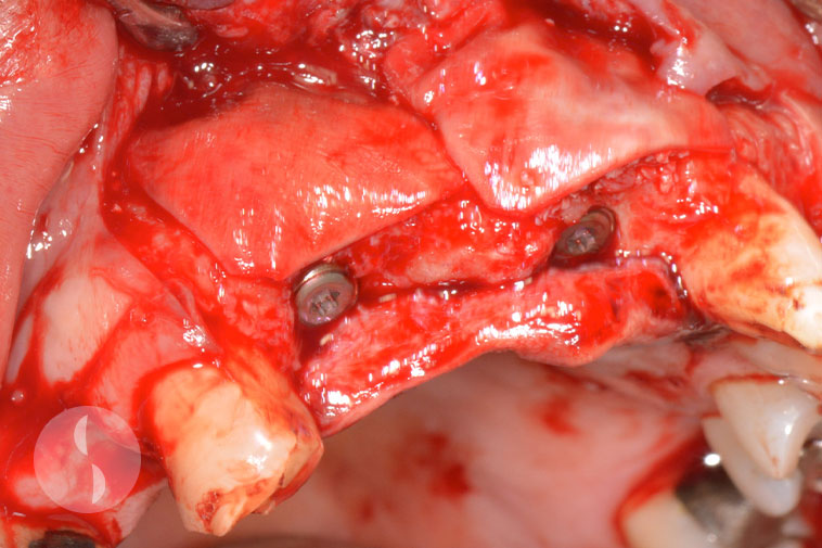
Guided bone regeneration to improve buccal profile
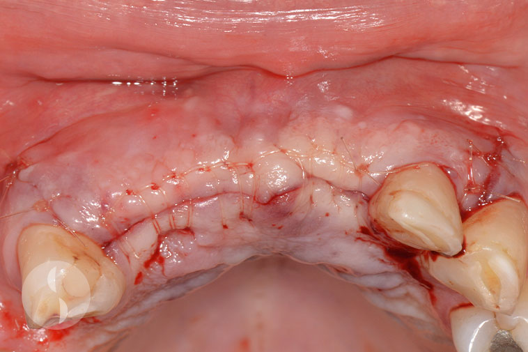
Immediately post operative
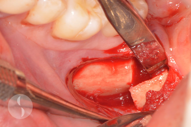
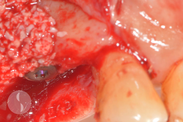
Bone Augmentation
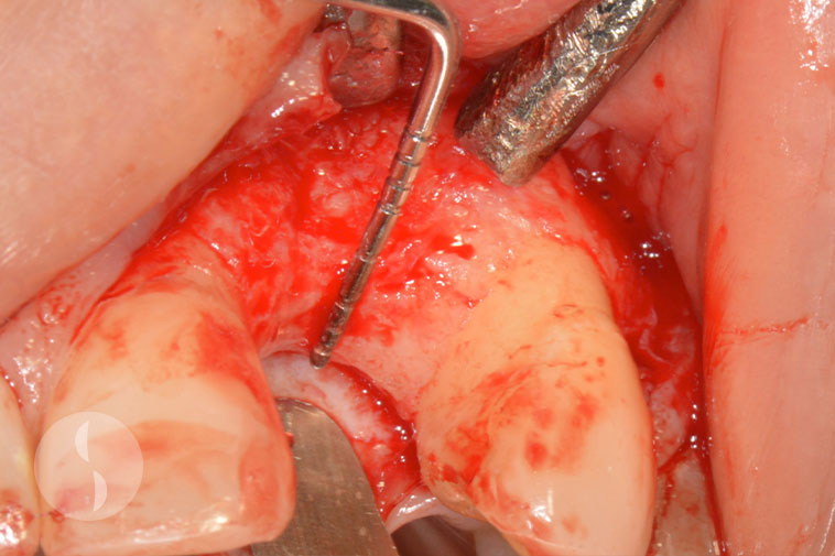
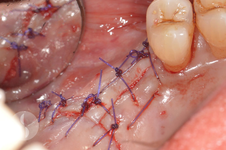
Case 1
Endodontic abscess resulting in large buccal defect
Particulate grafting following debridement of defect
Re-entry surgery after 3 months showing excellent integration of graft and remodeling
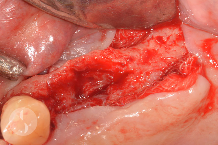
Case 2
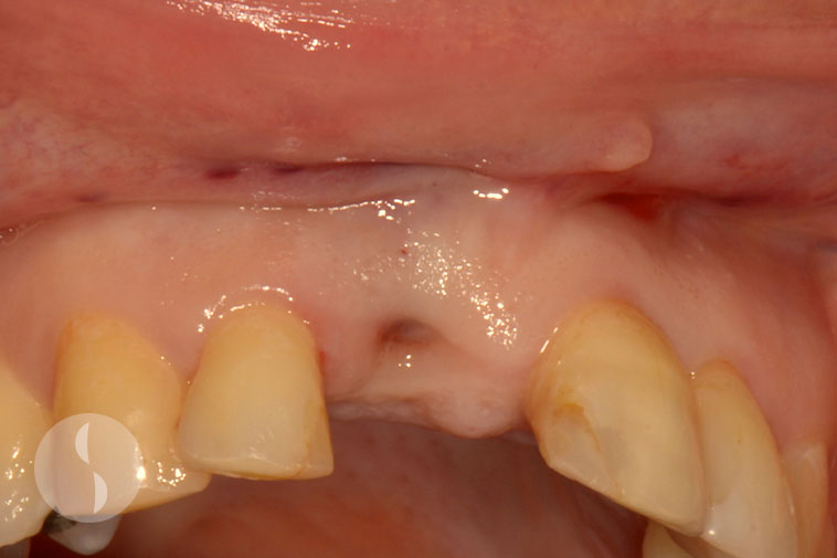
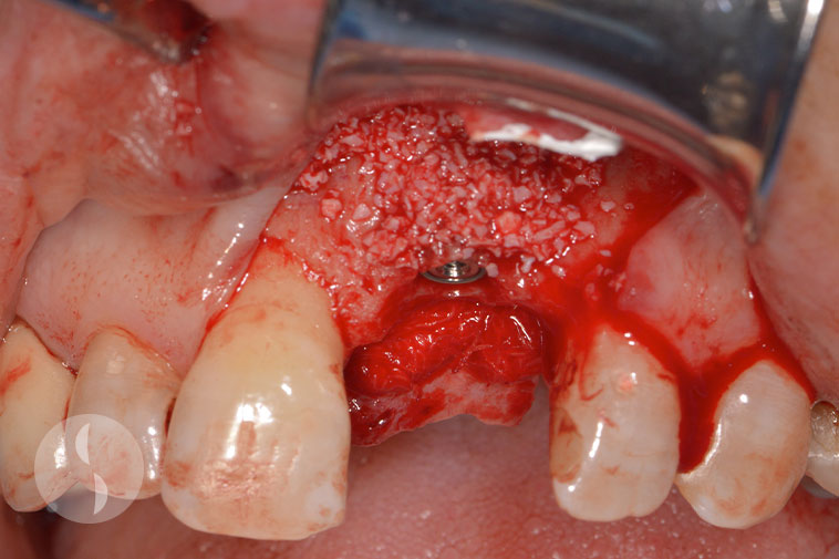
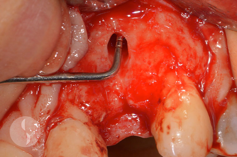
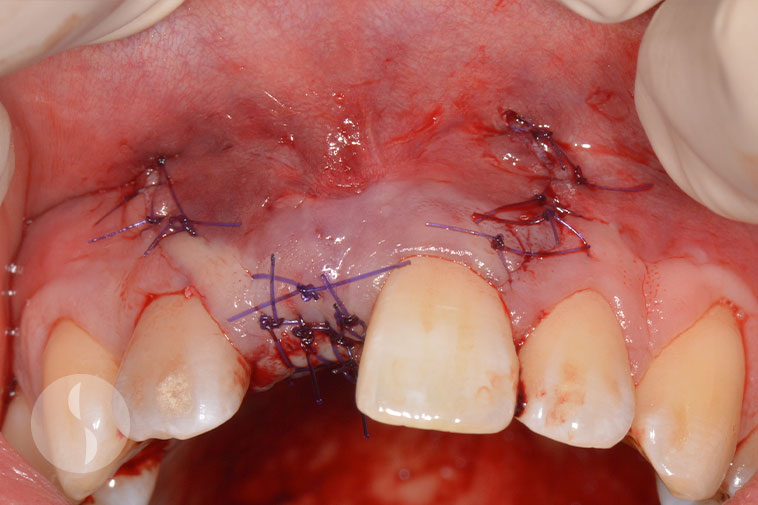
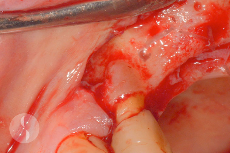
Preoperative situation showing missing 11
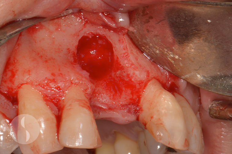
Large buccal defect due to endodontic abscess
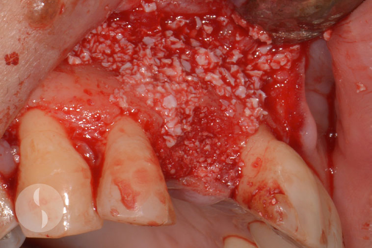
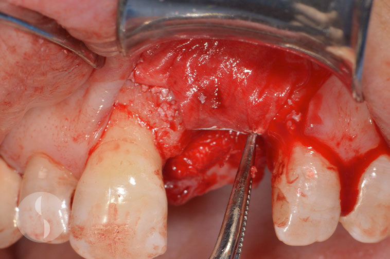
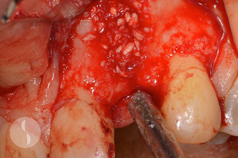
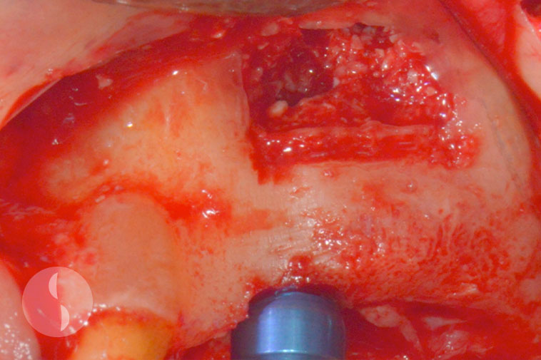
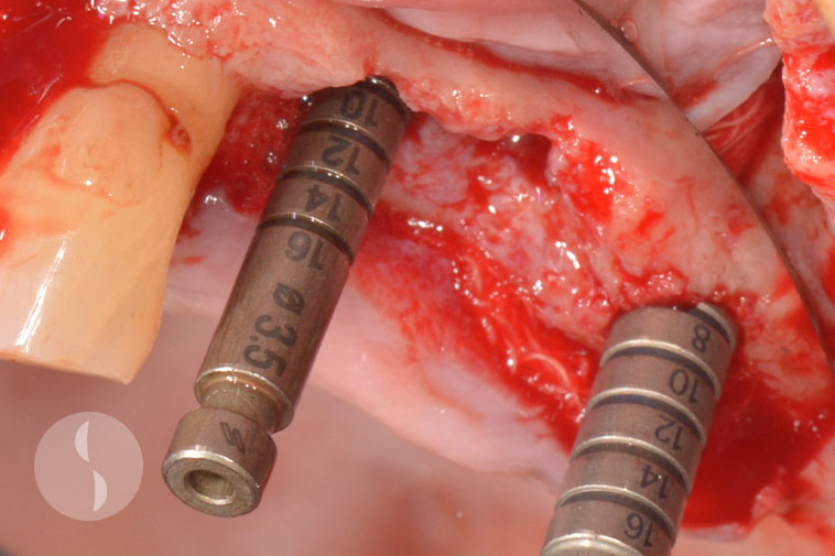
Placement of particulate graft (GBR)
Immediately post operative
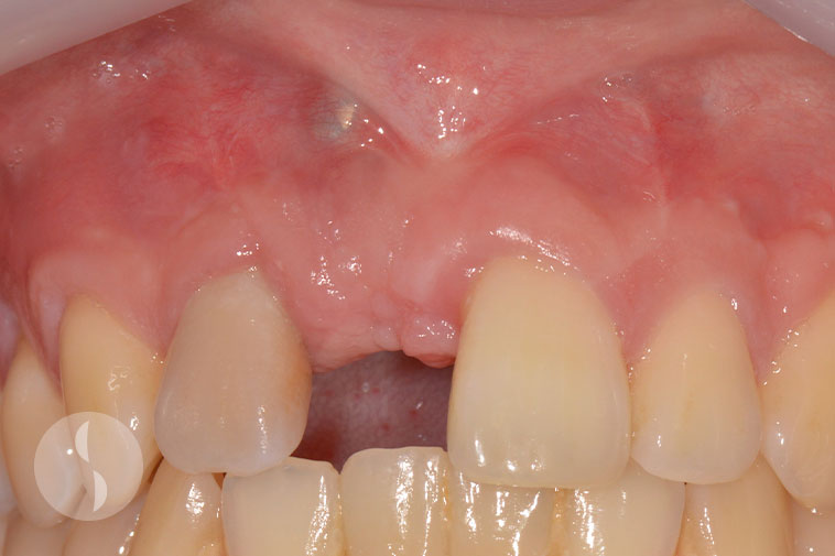
Implant placement in regenerated bone at 6 months following surgery
Case 3
Block graft harvested from mandible
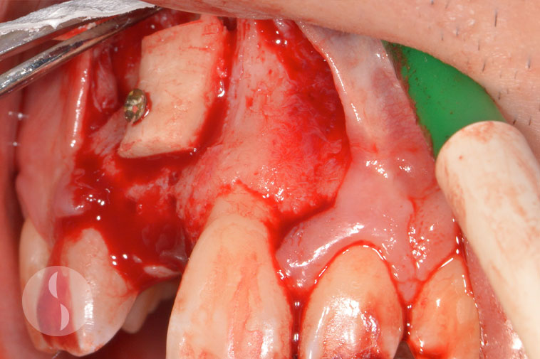
Block graft fixed to recipient site
Immediately post operative
Healing at 3 months following block graft surgery
Lateral Window Sinus
Case 1
Lateral window sinus lift procedure
Sinus grafted and implant placed
Immediately post operative showing implant placed with single stage technique
Soft tissue healing at 3 weeks following surgery
Transcrestal Sinus Lift
Case 1
Osteotome sinus lift at 16 site
Summer’s technique for transcrestal approach
Buccal graft to improve countour
Implant placed with submerged approach
Case 2
Edentulous ridge with socket defects
Implants placed in ideal prosthetic position
Defects grafted with GBR technique
Immediately post operative
Successful bony integration following sinus floor elevation
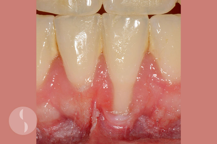
Soft Tissue Grafting
Case 1
Before
After
Case 2
Before
After
Case 3
Before
After